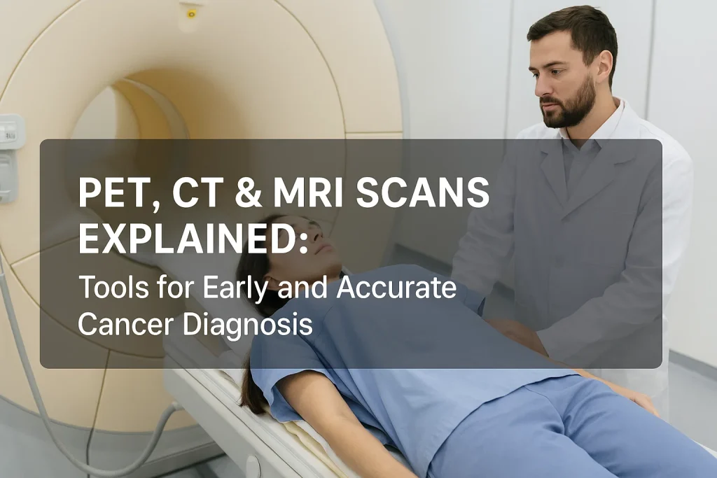
PET, CT & MRI Scans Explained: Tools for Early and Accurate Cancer Diagnosis
Cancer diagnosis is a complex process that depends mainly on advanced imaging techniques. The introduction of the Key tools in modern cancer management, including PET scans, CT scans, and MRIs, can help determine tumor size, location, spread, and response to treatment. Knowing the differences between PET, CT, and MRI scans—and understanding when to use each—is crucial for patients and caregivers throughout the cancer journey.
In this article, we will explain the types of scans, how each scan works, and how they help in early and accurate cancer diagnosis and monitoring.
The Importance of Imaging in Cancer Diagnosis
Medical imaging is essential for detecting, staging, and monitoring cancer. When someone shows symptoms or a doctor suspects cancer, imaging is often the first step toward a clear diagnosis. Whether it’s identifying tumors, analyzing their extent, or determining treatment response, scans like PET, CT, and MRI offer a non-invasive view inside the body.
What is the Role of Imaging in Cancer Diagnosis?
Medical imaging plays a crucial role in:
- Identifying tumors
- To determine the stage of cancer
- Monitoring treatment response
- Treatment plan for surgery and radiotherapy
- Detecting cancer recurrence
What are the three most common tools used in Cancer Diagnosis?
The three most commonly used tools in cancer diagnosis are
- Positron Emission Tomography (PET) scan
- Computed Tomography (CT) scan
- Magnetic Resonance Imaging (MRI)
Note: The use of diagnostic tools depends on the patient’s medical history and condition. Clinical correlation is crucial for confirming a diagnosis and guiding further treatment.
PET Scan: Monitoring cancer metabolism.
What is a PET Scan?
A PET scan (Positron Emission Tomography) is a nuclear imaging test that uses a radioactive tracer to monitor cellular activity throughout the body. Because cancer cells grow fast, they use more glucose than normal cells. PET scans identify these “hot spots,” where metabolic activity is higher.
How does a PET scan work?
Preparation and Procedure
- The patient is injected with a small amount of radioactive sugar (commonly FDG—fluorodeoxyglucose).
- The tracer moves through the body, and the amount of tracer absorbed by cells is directly proportional to cell metabolism.
- A PET scanner detects areas of high metabolic activity by emitting radiation and creates a 3D image that appears brighter on scans, often indicating cancer.
Key Features of PET Scan
- Helps detect cancer metabolism and spread
- Best for Lymphoma, lung cancer, colorectal cancer, and melanoma
Advantages of PET Scans in Cancer Detection and Treatment
- Detects cancer before any structural changes occur.
- Helps identify whether a tumor is cancerous or non-cancerous.
- Helps monitor treatment effectiveness, such as for chemotherapy or radiation therapy.
- Useful in detecting cancer relapse.
PET Scan Limitations
- Sometimes less effective in diagnosing Small Tumors.
- It may provide false results in case of infection or inflammation.
- Limited availability and often expensive.
CT scan: Detailed analysis of tumor structure
What is a CT Scan?
A CT (Computed Tomography) scan creates cross-sectional images of the body by utilizing X-ray technology. It’s similar to taking multiple X-ray images from various angles and combining them into a detailed 3D image.
CT scans are often the first imaging test recommended when cancer is suspected.
CT Scan Procedure
- The patient lies on a motorized table.
- The table moves into a large, doughnut-shaped scanner.
- X-rays rotate around the body, capturing images from different angles.
- A detailed 3D image is generated to check for any tumor structure.
CT scan key features
- Helpful in detecting masses and visualizing detailed structure
- Advised for lung, liver, kidney, and pancreatic cancers
Benefits of CT Scans
- Widely available, quick process
- Perfect for visualizing bone and organ structure.
- Helpful in planning treatments and staging tumors.
- Supports with surgeries and biopsies.
CT scan Limitation
- Uses Ionising radiation
- Less effective in soft tissues like the brain and spine.
- Might skip tiny tumors.
- Contrast dye (for high-resolution images) may cause allergies.
Note: Discuss with your doctor if you are sensitive to radiation and dye before a CT scan
MRI: For soft tissue Imaging
What is an MRI?
An MRI (Magnetic Resonance Imaging) scan uses powerful magnets and radio waves (rather than radiation) to create detailed images of soft tissues, organs, and internal structures. It’s especially effective on the brain, spinal cord, muscles, and connective tissues.
Procedure of an MRI scan
- The patient is lying in a big magnet-filled tube.
- The body’s hydrogen atoms align due to the magnetic field.
- Radio waves disrupt this alignment, and sensors detect the energy released when the atoms realign.
- A computer processes the signals into detailed images.
Preparation before the MRI scan
- Don’t wear any metallic objects or jewelry, as they may interfere with the detector’s magnetic waves, leading to inaccuracy in results.
- Keep yourself hydrated.
- If any additional preparation is required, take advice from your doctor
Key features of an MRI scan
- Helpful in soft tissue imaging.
- Best for brain, spinal cord, abdomen, breast, and prostate cancers.
- For higher resolution, a contrast may be required.
- Completed within 10-30 minutes.
Advantages of MRI
- Comes with no radiation.
- Detailed image quality for soft tissues.
- Helpful in diagnosing brain tumors and spinal metastasis.
- An advanced imaging technique is useful for breast cancer staging.
Limitations of MRI
- Long scan time.
- Not suitable for patients with metal implants or pacemakers.
- More costly and less available than CT.
- Motion-sensitive, requires stillness.
Comparison Table: PET vs CT vs MRI
| Criteria | PET Scan | CT Scan | MRI |
| Imaging type | Functional (metabolic) | Structural (anatomical) | Structural (soft tissues) |
| Radiation | Yes (uses low dose of radioactive tracer) | Yes (X-rays) | No (magnetic waves) |
| Time Taken | 30–60 min | 10–30 min | 30–60 min |
| Best For | Detecting spread, treatment response | Tumor size, location | Soft tissue detail |
| Common Use | Lymphoma, lung cancer | Liver, kidney, pancreas | Brain, spinal cord, breast |
| Cost | High | Moderate | High |
| Availability | Moderate | High | Moderate |
When Do Doctors Use PET, CT, or MRI Scans for Cancer?
1. Initial Diagnosis
- Most often, a CT scan is the first imaging test.
- If soft tissue or nervous system involvement is suspected, MRI is recommended.
- To monitor the cancer spread, A PET scan may be followed.
2. Staging
Making treatment decisions requires accurate staging:
- CT and MRI scans help with the location and size of tumors.
- A PET scan locates distant metastases.
3. Follow-up Treatment
- A PET scan is ideal for monitoring metabolic response.
- Tumor shrinkage is measured by a CT scan.
- MRI helps detect soft tissue tumors or brain metastases (spread).
4. Surveillance for Recurrence
- Post-treatment regular CT or MRI scans are used.
- If there is a suspicion of recurrence, PET scans are performed.
How Doctors Choose the Right Imaging Test for Cancer
The choice depends on:
- Cancer type
- tumor Location
- Stage of cancer
- Particular symptoms, if applicable
Most doctors use a combination of imaging tools; for example, A patient with lung cancer may undergo a CT scan to detect the tumor, a PET scan to assess spread, and an MRI if there is concern about brain metastases.
Risks & Safety of Imaging Procedures
While diagnostic imaging is generally safe, each has its own set of risks.
- Radiation Exposure: CT and PET use ionizing radiation. Repeated scans may slightly increase the risk of cancer, but the benefits usually compensate for the risks.
- Sensitivity to Contrast: CT and MRI scans may use contrast dyes, which can cause allergic reactions in some people.
- Anxiety and Discomfort: Some patients may find MRI scans to be stressful due to their length and noise.
Is Imaging Safe for Pregnant Women or Children?
Imaging during pregnancy is allowed under certain conditions. MRI is often safer because it uses no radiation, whereas CT and PET are avoided and handled with caution unless necessary.
Imaging doses for children are kept to a bare minimum. Pediatric oncologists ensure that scans are only performed when necessary.
How to be Prepared for Your Imaging Scan?
Understanding what to expect can ease anxiety:
- Fasting: You may be advised to avoid eating for 4–6 hours in case of PET scans.
- No metal objects: For MRIs, remove jewelry, watches, or anything metallic.
- Hydration: Drinking water helps flush out contrast material post-scan.
- Comfortable clothing: Be comfortable with your clothing unless changing into a gown is required.
Before going for a scan, inform your doctor. If you are pregnant, breastfeeding, or have kidney problems (especially for contrast-based scans),
Questions to Ask Your Oncologist
- What scan is needed, and why?
- What does the scan show?
- How much time will it take?
- Are there any adverse effects?
- Do I require a contrast dye?
- Is this scan covered by insurance?
Final Thoughts
The first step towards an accurate diagnosis is the right imaging. While PET scans show the metabolic activity of cancer before structural changes take place, MRIs offer unparalleled soft tissue contrast, and CT scans offer a rapid and detailed view of the body. Together, they provide a potent trio in the fight against cancer, with each scan providing particular information.
If you or a loved one is suspected of having cancer, ask your oncologist which scans are required and why. Knowing how these tools work will help you make correct decisions and feel more in control of the journey ahead.
FAQs
What’s the difference between a PET scan and a CT scan?
A PET scan shows metabolic activity, while A CT scan displays structural anatomy. PET is better for identifying active cancer cells; CT is better for anatomical detail.
Is MRI better than CT for cancer diagnosis?
Depending on the location of the cancer. MRI is better for the brain, spine, and soft tissue, while CT is more suitable for the lungs, liver, and bones. Discuss with your doctor before scanning.
Can these scans detect all types of cancer?
Depending on the stage and type, they can detect most types, but some early-stage cancers or microscopic tumors may require biopsies or additional tests to confirm.
Are Imaging scans painful?
No. These are painless procedures, though lying still for long durations may cause discomfort.
How often should cancer patients get these scans?
Your oncologist will decide the schedule based on cancer type, treatment plan, and risk of recurrence.
Do all cancer patients need all three scans?
No. The type, stage, and location of the cancer determine the kind and number of scans.
Can MRI, CT, or PET scans take the place of a biopsy?
No. Imaging scans can suggest whether a mass is unusual; to confirm this, a biopsy is preferred for cancer.
What happens if a scan finds something abnormal?
Further testing, like a biopsy, blood work, or additional imaging, will be scheduled to confirm or rule out cancer.
- Typhoid Fever: Symptoms, Causes, Diagnosis, Treatment Options, and Prevention Tips
- PCOS (Polycystic Ovary Syndrome): Causes, Symptoms, Diagnosis, and Treatment