CT Scans: Are They a Hidden Cancer Risk ?
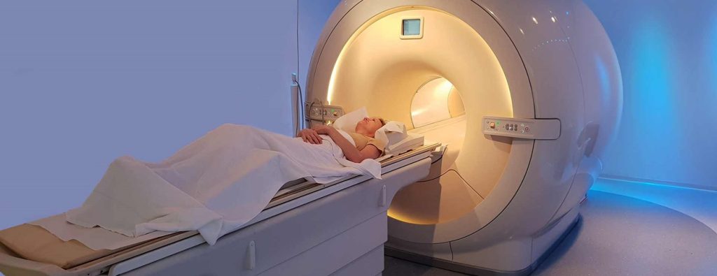
A CT scan, or computed tomography scan, is a diagnostic test that your doctor might use to identify issues inside your body, assist in surgical planning, or evaluate the effectiveness of treatments. The scan uses powerful X-rays, a form of radiation, to produce detailed images of the inside of your body.
Some people may be concerned about the radiation exposure from a CT scan, as radiation is known to potentially cause cancer. However, the risk of developing cancer from a CT scan is very low. For many, the benefits of detecting serious health conditions or monitoring treatment effectiveness outweigh this minimal risk.
How Do CT Scans Work?
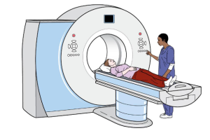
CT scans work by using a narrow X-ray beam that rotates around a specific part of your body. This rotation allows the machine to capture multiple images from various angles. These images are then processed by a computer to create a cross-sectional view of the area being examined. Much like a slice from a loaf of bread, this two-dimensional (2D) image shows a “slice” of your body’s interior.
The process is repeated multiple times, producing several slices. These slices are then stacked together by the computer to form a detailed, three-dimensional image of your organs, bones, or blood vessels. For instance, a surgeon might use this type of scan to examine all sides of a tumor to plan for surgery.

Radiation During a CT Scan
CT scans utilize X-rays, a form of ionizing radiation, which can potentially damage the DNA in your cells and increase the risk of cancer.
These scans expose you to more radiation than other imaging methods, such as standard X-rays or mammograms. For instance, a single chest CT scan can expose you to the same amount of radiation as 100 to 800 regular X-rays. Although this may seem significant, the overall amount of radiation you receive is still relatively low.
It’s important to remember that everyone is exposed to ionizing radiation daily from natural sources in the environment. On average, a person receives about 3 milli Sieverts (mSv) of radiation annually. A CT scan can deliver between 1 to 10 mSv, depending on the specific test and the area of the body being scanned. For example, a low-dose chest CT scan exposes you to approximately 1.5 mSv, while a standard-dose chest CT scan may expose you to around 8 mSv.
The more CT scans you undergo, the more radiation exposure you accumulate. However, this shouldn’t deter you from getting a CT scan if your doctor recommends it.
There are always concerns about radiation exposure from medical imaging. Patients frequently ask whether radiation from tests like mammograms, bone density tests, and CT scans will increase their cancer risk. For most women, the risk from routine X-ray imaging, such as mammograms or dental X-rays, is very low. However, many experts are worried about the growing use of higher-dose radiation tests, such as CT scans and nuclear imaging.
Over 80 million CT scans are performed annually in the United States, a significant increase from just three million in 1980. There are valid reasons behind this trend. CT scans and nuclear imaging have transformed the way we diagnose and treat medical conditions, reducing the need for exploratory surgeries and other invasive procedures that were once common and carried their own risks. When used appropriately, the benefits of these tests far outweigh the associated cancer risks, and the risk from a single CT scan or nuclear imaging test is quite small. However, we must consider whether this increasing reliance on such tests could lead to future public health concerns.

Will It Lead to Cancer?
What are the chances that the X-rays from a scan could cause a problem? It varies depending on your age, gender, and the part of your body being scanned. Generally, the risk is very low—the likelihood of developing a fatal cancer from any single CT scan is about 1 in 2,000.
Some organs are more vulnerable to radiation than others. Radiation tends to have a greater impact on cells that grow and divide rapidly. Therefore, organs like the breasts, lungs, thyroid gland, and bone marrow, which contain fast-dividing cells, are more sensitive to radiation than other parts of the body, such as the brain.
The risk of cancer from radiation is slightly higher in women than in men and is also higher in children. This is because children’s cells are dividing more rapidly as they grow, and they have more years ahead of them during which cancer could potentially develop.
How Can You Protect Yourself?
You don’t need to avoid CT scans altogether, but it’s wise to ensure that each one is necessary.
Before undergoing any imaging test, consider asking your doctor the following questions:
- Why do I need this scan?
- How will it impact my treatment?
- What are the risks involved?
- Is there a non-radiation alternative, like an MRI or ultrasound, that could be used instead?
- How will you protect the rest of my body during the CT scan?
Your doctor should aim to use the lowest possible radiation dose for the scan, especially if multiple scans are needed. Ask if the technician can shield other parts of your body with a lead apron to prevent unnecessary radiation exposure.
It’s also a good idea to keep track of all the CT scans and other X-rays you’ve had. This will help you monitor your cumulative radiation exposure and avoid unnecessary repeat tests. Record the following details:
- The type of scan
- The date it was performed
- The radiation dose received
- The name of the facility where the test was conducted

Exposure to Ionizing Radiation on the Rise
The radiation you receive from X-rays, CT scans, and nuclear imaging is known as ionizing radiation—high-energy wavelengths or particles that can penetrate tissues to visualize the body’s internal organs and structures. Ionizing radiation has the potential to damage DNA, and while your cells are capable of repairing most of this damage, they don’t always do so perfectly. This imperfect repair can leave behind small areas of “mis-repair,” leading to DNA mutations that may contribute to the development of cancer years later.
- Chemotherapy vs Radiation Therapy: Key Differences in Cancer Treatment
- Lung Cancer: Know the Risks, Take Action Now